Injury in medical practice is recognized as a traumatic lesion of tissues under the influence of certain negative mechanical factors, in which obvious external signs of damage are usually invisible.
Often, instead of the term "bruise," doctors use the concept of "concussion," which is one and the same.
Few can boast that he never received bruises: the notorious bruises, abrasions, bumps, these are just a few "companions" of the bruise.
These damages in the vast majority of cases do not pose any threat to health.
A completely different matter when it comes to eye injuries.
The human eye is a thin and fragile, but at the same time extremely important tool. Unlike most organs, it does not have sufficient protection (muscle corset, etc.) and is extremely vulnerable, but it is through it that we receive up to 90% of all information from the environment. At the same time, the eye is one of those organs that modern medicine has not yet studied to the end and, accordingly, many of the pathologies of the visual system have not yet learned to treat.
In order for the severity of eye contusion to be understood, it is enough to cite medical statistics. It is an eye injury in 85% of cases that causes the development of monocular blindness (blindness in one eye). At the same time, not everything is so sad, only in 5% of patients who received eye contusion, the outcome of the disease was a complete or predominant loss of vision in one eye.
It is precisely the reason for the danger of this eye injury that it is important to know what constitutes a bruised eye, what to do if trouble has already happened, and what should be avoided.
Based on the above general definition of a bruise, it can be formulated that a bruise of the eye is a traumatic damage to an organ caused by certain mechanical factors (direct physical impact, shock wave, etc.) that are not accompanied by a loss of eye integrity.
1. Eye injury: causes
The causes of bruises, as a rule, are of a domestic nature. One way or another, a bruise always provokes a mechanical effect. Most often, we are talking about a hit (hit the ball in the face during the game, hit during a fight or sparring, the impact of the shock wave from an explosion, etc.). No one is safe from injury, but children, athletes (especially when it comes to contact sports), military personnel, as well as those whose professional activities are associated with high physical activity, are most likely to suffer this type of injury.
The immediate cause of the injury is the transfer of kinetic energy from the body to the orbit and the eyeball. As a result of energy transfer, blood vessels rupture and tissue damage.
2. Eye injury: symptoms
The intensity of the symptoms, as well as its nature will vary from case to case. Two of these factors depend on:
A) the degree of physical impact (severity of injury);
B) the time elapsed since the injury.
The most common symptoms of eye contusion include:
1) Pain. The intensity of the pain syndrome, as well as its duration, is very individual. The strength of pain depends on the pain threshold, as well as on the severity of an eye injury. Be that as it may, a blow of any strength leads to a bruise, but it is far from always accompanied by pain. So, pain is one of the first symptoms, although in some cases it may be absent altogether.
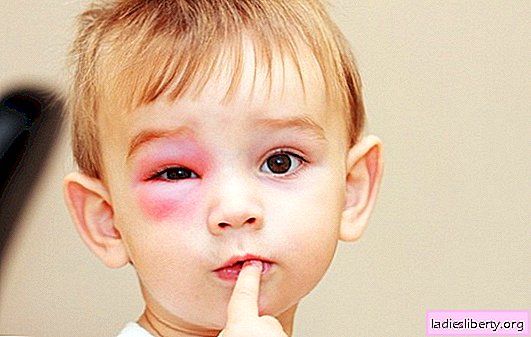
2) Swelling of the eyelids and periocular tissue. It develops with severe bruises. The causes of edema are the intense accumulation of fluid in places of tissue damage. Edema can form 8-12 hours after injury. In some cases, earlier.
3) Discomfort in the eye. The reason lies in the irritation of many nerve endings, and is also caused by proliferating edema.
4) "Fog" before the eyes, blurred perception. Visual impairment is an alarming symptom of eye contusion, as is nonspecific, which means that it can accompany everything, up to the developing retinal detachment.
5) Tear.
6) Fear of the light.
General symptoms may be present. Among the common symptoms are:
1) Increase in body temperature. The reason lies at the beginning of the inflammatory process in the area of damaged tissues.
2) Nausea, vomiting.
3) Dizziness.
Quite often, a bruise is not terrible in itself, but due to the fact that it entails severe violations in the operation of the visual analyzer. The most common:
• Retinal Damage. A bruise can lead to the formation of ruptures of the retina, up to its detachment. Also important to note. About 40% of myopic people have such a quiet, but extremely dangerous disease, such as retinal dystrophy. Peripheral retinal dystrophy leads to a thinning of the peripheral parts of the nerve membrane of the eye and increases the risk of ruptures with subsequent possible detachment. Often, peripheral retinal dystrophy is observed in people with normal vision. In people with dystrophy, which they most often do not suspect until the thunder strikes, the risk of damage to the retina increases significantly.
• Corneal damage. With significant bruises, the so-called traumatic cataract, or, otherwise, clouding of the lens. In addition, the beginning of the process of its destruction is possible.
• Damage to ligaments and oculomotor muscles. Another dangerous consequence that occurs as a result of eye contusion is damage to the ligaments responsible for the “tuning" of the lens. In this case, parallel damage to the lens and loss of transparency occurs.
• A bruise can cause an iris rupture. As a result, the pupil loses its ability to respond to light and expands.
• Bleeding into the eye chamber. Otherwise - hemophthalmus. The result is decreased vision, possible retinal detachment.
3. Eye injury: diagnosis
Diagnosis of eyeball contusion is not particularly difficult. In typical cases, the picture of the injury is obvious and a quick examination is determined even by an inexperienced ophthalmologist.
The most important task of a doctor is to eliminate the most dangerous consequences of a bruise (listed above). In order to exclude the following activities:
1. Fundus examination. The main mistake of ophthalmologists is the so-called fundus examination of the fundus with a narrowed pupil. Everyone has probably known an ophthalmoscope since childhood (a kind of mirror with a hole in the center).
Unfortunately, this method of examination of the fundus is not very informative and shows only the most gross changes on the part of the retina (its central region - the macula and the immediate periphery).
Apparently, about 65% of the retina remains, and if the study was carried out with a narrowed pupil - much more. A kind of "gold standard" of the study of the state of the retina is examination on a special ophthalmoscope with a Goldman lens. Thanks to him, you can examine even the most stressed areas of the retina.
"Private" polyclinics of the CIS countries are not equipped with such, so it is highly advisable to contact a specialized eye hospital, or insist on conducting a study with mydriasis (pupil dilation).
2. Visual acuity test. A standard check using a table allows you to determine visual acuity. With eye contusion, the decrease can be quite noticeable.
3. Computer perimetry, other types of perimetry. Allows you to determine the field of view. In case of serious damage, scotomas (loss of visual fields) are formed, which are precisely determined by perimetry.
Depending on the severity of the case, the list may be expanded or narrowed, at the discretion of the treating specialist.
Do not lose vigilance to the patient. You should know exactly the most threatening symptoms that you need to immediately contact a doctor:
• Flickering in the field of view of photopsies (bright stars, lines, zigzags, dots), as well as the appearance of other "artifacts" in the field of view.
• A sharp increase in the number of opacities in the eye (worms, stars, bunches and other floating opacities).
• Decreased lateral vision. To independently check the field of view, you can perform the simplest perimetry.
A) Fix the gaze at one point.
B) Extend your hand in front of you, extend your thumb (should be strictly at eye level).
C) Slowly move your hand with your finger to the side until the angle between the body and the hand becomes 90 degrees.
D) Determine whether the finger is visible. Since far peripheral vision is most sensitive to movement, several movements of the phalanx of the thumb can be made.
IMPORTANT! The background against which the thumb visibility will be evaluated should be contrasting (best of all - daylight from the window).
• The appearance in the field of view of a black dense or translucent curtain.
4. Eye injury: treatment
In most cases, special treatment is not required. It is important to monitor the general condition, as well as to note whether “dangerous” symptoms are occurring. However, if the injury is serious, the following can be prescribed:
• Supportive eye drops.
• Local antibacterial drugs (drops) to prevent the development of infection and inflammation.
Self-medication is not recommended. It is also not recommended to abuse folk remedies (an allergic reaction may develop).
Thus, eye injury, the injury is not so common, but very dangerous in its consequences. This is definitely not the case when going to the doctor can be neglected. There is a high risk of developing extremely severe consequences for vision, the worst of which is retinal detachment.
From the point of view of diagnosis, it is more important not to determine the injury, but to eliminate the occurrence of dangerous consequences. Only an experienced doctor can handle this.